Serena Bonaretti, PhD

I am a researcher at Balgrist Campus. My background is in medical image acquisition and analysis and biomechanics applied to the musculoskeletal system.
I am leading the creation of the Open and Reproducible Musculoskeletal Imaging Research (ORMIR) community, where we develop open and reproducible workflows to analyze musculoskeletal images.
I also promote transparent research on my YouTube channel and in talks and workshops.
Finally, I am writing the book Learn Python with Jupyter.


Complete CV: here

serena.bonaretti.research@gmail.com


Research Projects

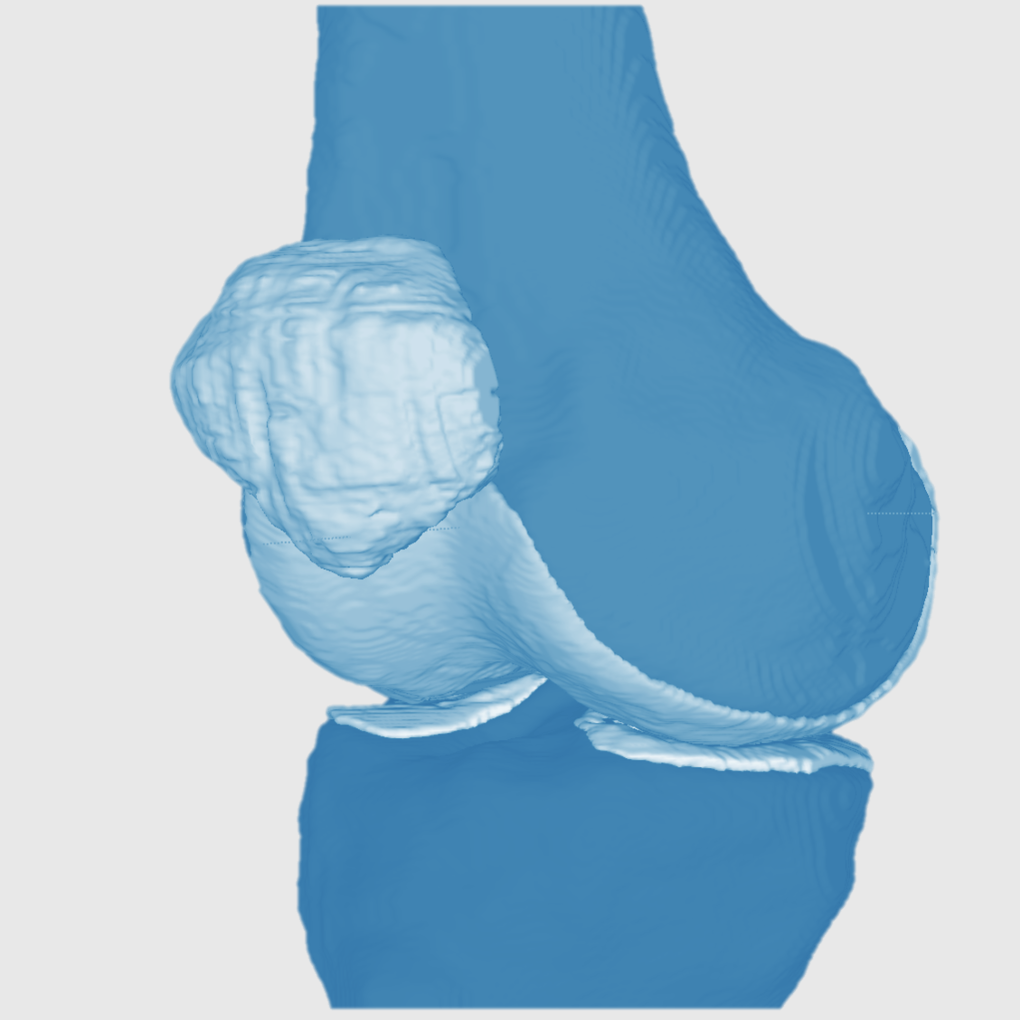
pyKNEEr: An Image Analysis Workflow for Open and Reproducible Research on Femoral Knee Cartilage

An automatic method to preprocess, segment, and analyze femoral knee cartilage for transparent research
PIs: Gary Beaupre (VA Palo Alto, Stanford University) and Garry Gold (Stanford University)
Code: Zenodo
Documentation:
Publications:
  • pyKNEEr: An Image Analysis Workflow for Open and Reproducible Research on Femoral Knee Cartilage. PLoS ONE 15(1): e0226501. 2020. .bib
  • pyKNEEr: Reproducible Workflow for Automatic Segmentation and Analysis of Femoral Knee Cartilage. QMSKI 2019. 24 February - 1 March 2019. Chateau Lake Louise, Canada.


Weight-Bearing Imaging of the Knee Using C-Arm CT

Development of a novel weight-bearing C-Arm CT system to expand understanding of cartilage mechanical stresses and to provide quantitative measures of knee joint health.
PI: Garry Gold (Stanford University)
Publications:
  • Comparison of Different Approaches for Measuring Tibial Cartilage Thickness. J Integr Bioinform. 14(2),1-10. 2017. .bib
  • Feasibility of Motion Compensation using Intertial Measurements in C-arm CT. IEEE Nuclear Science Symposium & Medical Imaging Conference. 10-17 November 2018. Sydney, Australia.
  • Object Removal in Gradient Domain of Cone-Beam CT Projections. IEEE Nuclear Science Symposium & Medical Imaging Conference. 29 October – 5 November 2016. Strasbourg, France.


Standardization of HR-pQCT for Bone Quality Assessment

In multicenter studies, standardization of acquisition procedures is fundamental for data comparison. In this project, we aimed to standardize the definition of the volume of interest and the measurement of bone mineral density.
PI: Andrew J. Burghardt (UCSF)
Web application:
Publications:
  • Operator Variability In Scan Positioning is a Major Component of HR-pQCT Precision Error and is Reduced by Standardized Training. Osteoporos Int. 28(1), 245-257. 2017. .bib
  • The Comparability of HR-pQCT Bone Quality Measures Is Improved by Scanning Anatomically Standardized Regions. Osteoporos Int. 28(7), 2115-2128. 2017. .bib
  • Statistical Parametric Mapping of HR-pQCT Images: A Tool for Population-Based Comparison of Micro-Scale Bone Features. Ann Biomed Eng. 45(5), 949-962. 2017. .bib
  • Quantifying Sex, Race and Age Specific Differences in Bone Microstructure Requires Measurement of Anatomically Equivalent Regions. Bone. 101, 206-213. 2017. .bib
  • Standardized Training For HR-pQCT Scan Positioning Reduces Inter-Operator Precision Errors: The MrOS Multicenter Study Experience. ASBMR 2015. 9-12 October 2015. Seattle, WA, USA.
  • The Role of Intra- and Inter-Operator Variability in HR-pQCT Precision. IBDW 2014. 13-17 October 2014. Hong Kong.
  • Intra- and Inter-Operator Variability in HR-pQCT Scan Positioning. ASBMR 2014. 12-15 September 2014. Houston, TX, USA.
  • Comparability of HR-pQCT Bone Quality Measures Improved by Scanning Anatomically Standardized Regions. ASBMR 2014. 12-15 September 2014. Houston, TX, USA.
  • Automated Scan Prescription For HR-pQCT: A Multi-Atlas Prospective Registration Approach. ASBMR 2013. 4-7 October 2013. Baltimore, MD, USA.


Standardization of QCT for Bone Quality Assessment

QCT inter-scanner variability is still a big challenge. We aimed to find cross-calibration laws using a novel anthropomorphic hip phantom.
PI: Thomas Lang (UCSF)
Publications:
  • Novel Anthropomorphic Hip Phantom Corrects Systemic Interscanner Differences in Proximal Femoral vBMD. Phys Med Biol. 59(24), 7819-34. 2014. .bib
  • Automatic Multi-Parametric Quantification of the Proximal Femur with QCT. Quant Imaging Med Surg. 5(4), 552-68. 2015. .bib
  • Inter-scanner Differences in In Vivo QCT Measurements of the Density and Strength of the Proximal Femur Remain After Correction with Anthropomorphic Standardization Phantoms. Med Eng and Phys. 36(10), 1225-32. 2014. .bib
  • QCT Intra- and Inter-Scanner Precision In Estimation Of Proximal Femur Strength. ASBMR 2015. 9-12 October 2015. Seattle, WA, USA.
  • Effect of Body Size on the Quantification of Bone Mineral Density From QCT Images Using a Novel Anthropomorphic Hip Phantom. ASBMR 2013. 4-7 October 2013. Baltimore, MD, USA.


Statistical Appearance Models for Bone Mechanics

My PhD project aimed to create a statistical appearance model of femur for finite element simulations of different populations.
PIs: Mauricio Reyes and Philippe Büchler (ISTB, Switzerland)
Code:
Publications:
  • Image-based vs. Mesh-based Statistical Appearance Model of the Human Femur: Implications for Finite Element Simulations. Medical Engineering and Physics. Dec;36(12):1626-35. 2014. .bib
  • The Virtual Skeleton Database: An Open Access Repository for Biomedical Research and Collaboration. J Med Internet Res. 12;15(11):e245. 2013. .bib
  • Evidence Based Development of a Novel Lateral Fibula Plate (VariAx Fibula) Using a Real CT Bone Data Based Optimization Process During Device Development. Open Orthop J. 6,1-7. .bib
  • Mesh-based vs. Image-based Statistical Model of Appearance of the Human Femur: a Preliminary Comparison Study for the Creation of Finite Element Meshes. Mesh Processing in Medical Image Analysis - MICCAI 2011 workshop. 18 September 2011. Toronto, Canada.
  • Combined Statistical Model of Bone Shape and Mechanical Properties for Bone Modelling. 9th International Symposium on Computer Methods in Biomechanics and Biomedical Engineering. 24-27 February 2010. Valencia, Spain.
  • Assessment of Peri-Articular Implant Fitting Based on Statistical Finite Element Modelling. Computational Biomechanics for Medicine III – MICCAI 2008 workshop. 10 September 2008. New York, NY.
  • Methods to Accelerate Finite Element Calculations in Biomechanics Using a Statistical Database of Pre-Calculated Simulations. 10th International Symposium on Computer Methods in Biomechanics and Biomedical Engineering. 11-14 April 2012. Berlin, Germany.
  • Statistical Model of Appearance to Accelerate Finite Element Calculations in Biomechanics. 19th Congress of the European Society of Biomechanics. 1-4 July 2012. Lisbon, Portugal.
  • Statistical Finite Element Model for the Virtual Skeleton Database. NCCR Co-Me Scientific Advisory Board Meeting. 9-10 February 2011. Interlaken, Switzerland.
  • Statistical Finite Element Modeling: Application to Orthopaedic Implant Design. Graduate School for Cellular and Biomedical Sciences Symposium. 28 January 2011. Bern, Switzerland.
  • Statistical Finite Element Modeling for the Virtual Skeleton Database. NCCR Co-Me Research Networking Workshop. 26-27 August 2010. Zürich, Switzerland.
  • A Statistical Shape Model of Bone Anatomical Variability for Finite Element Assessment of Bone Mechanics. 17th Congress of the European Society of Biomechanics. 5-8 July 2010. Edinburgh, Scotland.
  • Statistical Finite Element Modeling for the Virtual Skeleton Database. NCCR Co-Me Scientific Advisory Board Meeting. 19-20 February 2010. Winterthur, Switzerland.
  • A Statistical Shape Model of Bone Anatomical Variability for Finite Element Assessment of Bone Mechanics. Graduate School for Cellular and Biomedical Sciences Symposium. 27 January 2010. Bern, Switzerland.
  • Computing Average Anatomical Images: Comparison between Thin-Plate Spline and Log-Euclidean Approach. The Annual Meeting of the Swiss Society for Biomedical Engineering. 27-28 August 2009. Bern, Switzerland.
  • Automatic Bone Density Evaluation from CT Images. Computer Assisted Orthopaedic Surgery. 17-20 June 2009. Boston, MA.
  • Shape and Biomechanical Model for Population-Specific Design of Anatomical Peri-Articular Implants. CTI Medtech Event. 2 September 2008. Bern, Switzerland.
  • Statistical Finite Element Analysis for Bone Modelling. 16th Congress of the European Society of Biomechanics. 6-9 July 2008. Lucerne, Switzerland.
  • Statistical Finite Element Model for Bone and Implant Modeling. NCCR Co-Me Scientific Advisory Board Meeting. 14 February 2008. Neuchatel, Switzerland.
  • Statistical Models of Shape and Density for Population-based Analysis of Bone Mechanics with Applications to Fracture Risk Assessment and Implant Design. Ph.D. thesis. 2011.

Open Source Code

My own projects

pyKNEEr: An image analysis workflow for open and reproducible research on femoral knee cartilage

An automatic method to preprocess, segment, and analyze femoral knee cartilage for transparent research.

Language: Python with Jupyter Notebooks, SimpleITK, and Elastix
Code repositories: and Zenodo
Data repository:
Documentation:
Video:
Publication:

Statistical Appearance Models for Bone Mechanics

Statistical appearance model of femur for finite element simulations of different populations.

Language: C++ with ITK , VTK , and Qt
Repository:
Publication:

Supervised projects

Reference Line - Training and Evaluation

A webapp to train and evaluate HR-pQCT operators to acquire standardized images.

Developer: Caroline Mai Chan
Co-supervisor: Andrew Burghardt
Webapp:
Language: HTML with CSS and Javascript
Repository:
Publication:

FEM Assigner

A software to apply bone material properties to FE mesh from QCT images.

Developer: Andreas Siegrist
Co-supervisor: Benedikt Helgason
Language: C++ with ITK , VTK , and Qt
Repository:

Teaching

PYTHON TEACHING

Python language and scientific Python (Jupyter Notebook, Numpy, Pandas, Matplotlib, Keras, etc.) at C as in Coding.
I am collecting the content of my course into a book called Learn Python with Jupyter.


ONLINE TEACHING

YouTube channel with basics and hands-on tutorials on Open and Reproducible Research:


    • ACADEMIC TEACHING

      Surgery without all the blood (RAD 70N)

      2017. Guest Lecturer. Stanford University, USA

      Introduction to Interventional Radiology at the Zeego Lab (Demo class)


      Orthopaedic Bioengineering (BIOE/ME 381)

      2017. Co-instructor 50%. Stanford University, USA

      • Bone Anatomy and Physiology
      • Bone Mechanics I
      • Bone Mechanics II
      • Bone Imaging I
      • Bone Imaging II
      • Bone Quality and Fracture


      Clinical Needs and Technology (BIOE 301B)

      2017, 2018. Guest Lecturer. Stanford University, USA

      • Introduction to X-ray-based Imaging
      • Minimally Invasive Therapies in Swine (Laboratory)


      Introduction to Bioengineering Research (BIOE 390 / MED 289)

      2016. Guest Lecturer. Stanford University, USA

      Weight-bearing Imaging of the Knee Using C-arm CT (Presented project still under development)


      Image Processing and Analysis II (BI 265)

      2015. Guest Lecturer. University of California San Francisco, USA

      Active Shape and Appearance Modeling in Medical Imaging (Blackboard class)


      Musculoskeletal Imaging (BI 240)

      2014, 2015. Guest Lecturer. University of California San Francisco, USA

      Assessment of Bone Strength - Foundations of FE and microFE (Blackboard class)


      Medical Image Analysis

      2009. Guest Lecturer. ETH Zürich and University of Bern, Switzerland

      Statistical Shape Models